2023-09-26 22:35
UCLA Team Enhances Nobel Prize-Winning Tech
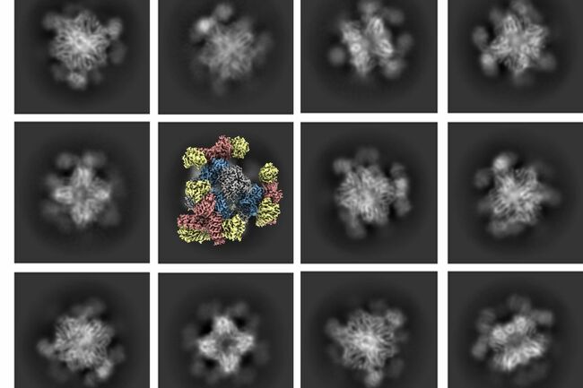
Roger Castells-Graells/UCLA A series of cryo-EM images. The greyscale photos represent 2D projections of multiple views of the imaging scaffold attached to a target protein; the color image illustrates the 3D reconstruction derived from 2D projections. Key takeaways A technology called cryo-electron microscopy, or cryo-EM, enables scientists to see the atomic structure of biological molecules in high resolution. But to date, it has been ineffective for imaging so-called small molecules. A UCLA-led team of biochemists devised a solution that makes it possible to hold small protein molecules in place while they're being imaged, which will enable cryo-EM to produce much clearer images of such molecules.
https://www.miragenews.com/ucla-team-enhances-nobel-prize-winning-tech-1092328/
#miragenews
https://www.miragenews.com/ucla-team-enhances-nobel-prize-winning-tech-1092328/
#miragenews
Sinun täytyy kirjautua sisään ennen kuin voit kommentoida.